


Page 187 - TBE_Book_V2_2019
P. 187
Chapter 10: Diagnosis


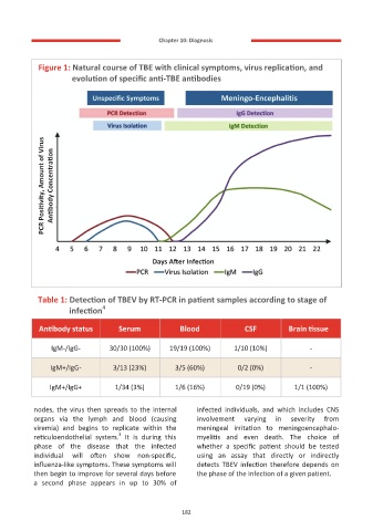
              Figure 1: Natural course of TBE with clinical symptoms, virus replication, and
                    evolution of specific anti-TBE antibodies


             PCR Positivity, Amount of Virus  Antibody Concentration


                                          Days After Infection


            Table 1: Detection of TBEV by RT-PCR in patient samples according to stage of
                            4
                    infection
           Antibody status       Serum           Blood            CSF         Brain tissue


               IgM-/IgG-      30/30 (100%)    19/19 (100%)      1/10 (10%)          -

               IgM+/IgG-       3/13 (23%)       3/5 (60%)        0/2 (0%)           -

              IgM+/IgG+         1/34 (3%)       1/6 (16%)       0/19 (0%)       1/1 (100%)


          nodes, the virus then spreads to the internal   infected  individuals,  and  which  includes  CNS
          organs  via  the  lymph  and  blood  (causing   involvement   varying   in   severity   from
          viremia)  and  begins  to  replicate  within  the   meningeal  irritation  to  meningoencephalo-
                                 3
          reticuloendothelial  system.   It  is  during  this   myelitis  and  even  death.  The  choice  of
          phase  of  the  disease  that  the  infected   whether  a  specific  patient  should  be  tested
          individual  will  often  show  non-specific,   using  an  assay  that  directly  or  indirectly
          influenza-like symptoms. These symptoms will   detects  TBEV  infection  therefore  depends  on
          then begin to improve for several days before   the phase of the infection of a given patient.
          a  second  phase  appears  in  up  to  30%  of


                                                  182
                                                  182

