


Page 55 - TBE_Book_V2_2019
P. 55
Chapter 2b: The molecular and antigenic structure of TBEV


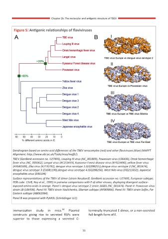
            Figure 5: Antigenic relationships of flaviviruses


         Dendrogram based on amino acid differences of the TBEV serocomplex (red) and other flaviviruses (blue) (MAFFT
         Alignment: http://www.ebi.ac.uk/Tools/msa/mafft/).
         TBEV (GenBank accession no. U27495), Louping Ill virus (NC_001809), Powassan virus (L06436), Omsk hemorrhagic
         fever virus (NC_005062), Langat virus (AF253419), Kyasanur Forest disease virus (AY323490), yellow fever virus
         (AY640589), Zika virus (KJ776791), dengue virus serotype 1 (GQ398255),dengue virus serotype 2 (NC_001474),
         dengue virus serotype 3 (EU081190),dengue virus serotype 4 (GQ398256), West Nile virus (DQ211652), Japanese
         encephalitis virus (D90194)
         Surface representations of the TBEV sE dimer (strain Neudoerfl, GenBank accession no. U27495, European subtype;
         PDB code: 1SVB, Rey et al., 1995) in pairwise comparisons with E of other viruses, displaying divergent surface-
         exposed amino acids in orange. Panel I: dengue virus serotype 2 strain 16681 (NC_001474). Panel II: Powassan virus
         strain LB (L06436). Panel III: TBEV strain Vasilchenko, Siberian subtype (AF069066). Panel IV: TBEV strain Sofjin, Far
         Eastern subtype (AB062064).
         Panel B was prepared with PyMOL (Schrödinger LLC).


                                      62
         immunization   study   in   mice.    Plasmid   terminally truncated E dimer, or a non-secreted
         constructs  giving  rise  to  secreted  RSPs  were   full-length form of E.
         superior  to  those  expressing  a  secreted  C-


                                                  50

