


Page 54 - TBE_Book_V2_2019
P. 54
Chapter 2b: The molecular and antigenic structure of TBEV


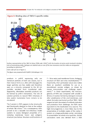
          Figure 4: Binding sites of TBEV E-specific mAbs


                                                      1
         Surface representation of the TBEV sE dimer [PDB code: 1SVB, ] with the location of amino acids involved in binding
         sites of neutralizing mAbs. Epitopes are labeled only on one of the two monomers and the mAbs are designated
         according to references. 92,95
         Color code of E as in Figure 1.
         The figure was prepared with PyMOL (Schrödinger LLC).


         products  in  prM-E  expressing  cells  are   E – Virus entry and membrane fusion; Antigenic
         immature  particles  of  both  size  classes,  but  in   structure of TBEV and virus neutralization). 54-60
         their mature forms (i.e. after prM cleavage) the   Importantly, their particulate nature also makes
         larger  particles  are  less  stable  and  therefore   them  an  excellent  candidate  for  use  as  a
         seen  as  a  minority  compared  to  the  30  nm   recombinant  vaccine  antigen,  as  shown  by
         particles  secreted  from  transfected  cells.   mouse  immunization  and  challenge  experi-
                                                           61
         Apparently,  alternative  assembly  products  can   ments.   In  these  experiments,  the  immuno-
         be  formed  by  prM-E  interactions.  The  role  of   genicity  of  RSPs  was  compared  with  soluble  E
         subviral  particles  in  natural  TBEV  infections  of   dimers, E rosettes formed by detergent removal
         ticks  and/or  mammalian  hosts  remains  to  be   after solubilization of the viral membrane, and
         elucidated.                                  whole formalin-inactivated purified TBEV. With
                                                      respect to both the extent of antibody induction
         The E protein in RSPs appears to be structurally   and  protection  from  challenge,  the  RSPs  were
         and functionally identical to that at the surface   equivalent to the inactivated virus vaccine. This
                          48
         of whole TBE virions.  As a consequence, RSPs   high  immunogenicity  is  most  likely  due  to  the
         proved  to  be  a  valuable  non-infectious  model   presentation of multiple copies of the native E
         systems  to  assess  biological  properties  of  E,   protein on a large particulate carrier, mimicking
         including  membrane  fusion  and  antigenic   its presentation on whole virus particles. Similar
         structure (see below: Structure and functions of   conclusions  were  also  derived  from  a  DNA

                                                  49

