


Page 49 - TBE_Book_V2_2019
P. 49
Chapter 2b: The molecular and antigenic structure of TBEV


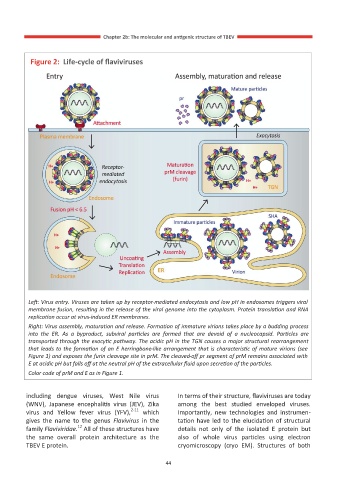
          Figure 2:  Life-cycle of flaviviruses


         Left: Virus entry. Viruses are taken up by receptor-mediated endocytosis and low pH in endosomes triggers viral
         membrane fusion, resulting in the release of the viral genome into the cytoplasm. Protein translation and RNA
         replication occur at virus-induced ER membranes.
         Right: Virus assembly, maturation and release. Formation of immature virions takes place by a budding process
         into  the  ER.  As  a  byproduct,  subviral  particles  are  formed  that  are  devoid  of  a  nucleocapsid.  Particles  are
         transported through the exocytic pathway. The acidic pH in the TGN causes a major structural rearrangement
         that leads to the formation of an E herringbone-like arrangement that is characteristic of mature virions (see
         Figure 1) and exposes the furin cleavage site in prM. The cleaved-off pr segment of prM remains associated with
         E at acidic pH but falls off at the neutral pH of the extracellular fluid upon secretion of the particles.
         Color code of prM and E as in Figure 1.


         including  dengue  viruses,  West  Nile  virus   In terms of their structure, flaviviruses are today
         (WNV),  Japanese  encephalitis  virus  (JEV),  Zika   among  the  best  studied  enveloped  viruses.
         virus  and  Yellow  fever  virus  (YFV), 2-11   which   Importantly,  new  technologies  and  instrumen-
         gives  the  name  to  the  genus  Flavivirus  in  the   tation have led to the elucidation of structural
                        12
         family Flaviviridae.  All of these structures have   details  not  only  of  the  isolated  E  protein  but
         the  same  overall  protein  architecture  as  the   also  of  whole  virus  particles  using  electron
         TBEV E protein.                              cryomicroscopy  (cryo  EM).  Structures  of  both

                                                  44

