


Page 48 - TBE_Book_V2_2019
P. 48
Chapter 2b: The molecular and antigenic structure of TBEV


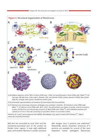
          Figure 1: Structural organization of flaviviruses


                                                                                       26
         (A,B) Ribbon diagrams of the TBEV sE dimer [PDB code: 1SVB, (1)] and full-length E dimer [PDB code: 5O6A, ]. (A)
             Top view. (B) Side view. Color code E: domain I (DI), red; domain II (DII), yellow; domain III (DIII), blue; fusion
             loop (FL), orange; stem, green; membrane anchor, grey.
         (C,D) Schematic representations of immature (C) and mature (D) virus particles.
         (E,F) Electron cryo-microscopy structures of dengue virus serotype 1 particles. (E) Immature virion [PDB code:
                  21
             4B03,  ]. (F) Mature virion [PDB code: 4CCT, (21)]. The prM proteins are shown in purple, and the E proteins
                                                                  26
             in gray. (G) Electron cryo-microscopy structure of TBEV [PDB code: 5O6A, ], with individual domains of E
             colored as in A to D. One raft consisting of 3 parallel E dimers is encircled in white.
                                                                               119
         Panels A, B, E and F were prepared with PyMOL (Schrödinger LLC), panel G with UCSF Chimera [ , http://
         www.rbvi.ucsf.edu/chimera/].

                                                                                            2
         DIII) that are connected to each other and the   (the  dengue  virus  E  protein)  was  published.
         membrane-associated  part  of  the  protein  by   Meanwhile,  atomic  resolution  structures  of  E
         flexible  linker  regions.  It  took  eight  additional   proteins  are  available  for  several  of  the  most
         years until another flavivirus E protein structure   important  human  pathogenic  flaviviruses,

                                                  43
                                                  43

