


Page 53 - TBE_Book_V2_2019
P. 53
Chapter 2b: The molecular and antigenic structure of TBEV


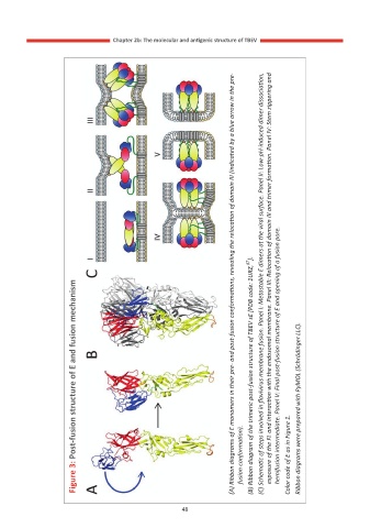
                                                                (A) Ribbon diagrams of E monomers in their pre- and post-fusion conformations, revealing the relocation of domain III (indicated by a blue arrow in the pre-


                      Figure 3: Post-fusion structure of E and fusion mechanism


                                                                  fusion conformation).  (B) Ribbon diagram of the trimeric post-fusion structure of TBEV sE [PDB code: 1URZ, 87 ].  (C) Schematic of steps involved in flavivirus membrane fusion. Panel I: Metastable E dimers at the viral surface. Panel II: Low-pH-induced dimer dissociation,  exposure of the FL and interaction with the endosomal m


                                                  48

