


Page 94 - TBE_Book_V2_2019
P. 94
Chapter 4: Pathogenesis of TBE


          significantly  increased  dose-dependent  TBEV   the  appearance  of  activated  caspase  3
          production,  whereas  the  inhibition  of   following  infection, and apoptosis in neurons
          autophagy   showed   a   dose-dependent     occurs in the same areas where viral antigen is
                                 11
          decrease of infectious virus.               present. 15,16   In  vitro,  TBEV  infection  causes
                                                      apoptosis  in  mouse  and  human  neural
          Apoptosis                                   cells. 17,18   Although  brain-infiltrating  CD8+  T
                                                      cells  contribute  to  the  fatal  outcome  during
          Apoptosis  is  a  process  of  programmed  cell    19
                                                      infection  no significant increase of apoptotic
          death  in  which  cells  activate  intracellular   cell death was detectable upon infection with
                        12
          death pathways.  This mechanism occurs in a   Langat  virus  (LGTV)  and  TBEV  in  mice. 5,20
          wide range of human viral infections, including   These data are in line with human data, where
          infections of the CNS  such as herpes simplex
                                                      no  prominent  signs  of  neuronal  apoptosis
          virus  (HSV)  and  cytomegalovirus  (CMV)   were  seen  in  post-mortem  brain  tissue  from
          encephalitis. 13,14   In  WNV  infection  of  mice,   patients.
                                                             21
          high virus titers in the CNS are associated with

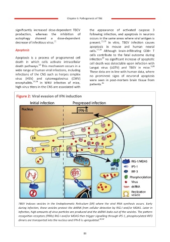
           Figure 2: Viral evasion of IFN induction


            TBEV  induces  vesicles  in  the  Endoplasmatic  Reticulum  (ER)  where  the  viral  RNA  synthesis  occurs.  Early
            during infection, these vesicles protect the dsRNA from cellular detection by RIG-I and/or MDA5. Later in
            infection, high amounts of virus particles are produced and the dsRNA leaks out of the vesicles. The pattern
            recognition receptors (PRRs) RIG-I and/or MDA5 then trigger signalling through IPS-1, phosphorylated IRF3
            dimers are transported into the nucleus and IFN-β is upregulated. 28,38


                                                     89

