


Page 95 - TBE_Book_V2_2019
P. 95
Chapter 4: Pathogenesis of TBE


          Type I IFN response                         most  studied.  Type  I  IFNs  binds  to  the  IFNα
                                                      receptor (IFNAR), which is expressed on nearly
          The  type  I  IFN  system  is  the  first  line  of   all  cell  types,  and  reacts  in  a  paracrine  and
          defense  against  viral  infection  and  an   autocrine manner. The IFNAR is composed of
          important part of the intrinsic innate immune   a  heterodimer  of  IFNAR1  and  IFNAR2.  After
          response that controls virus dissemination and   binding of IFN, the IFNAR activates the Janus
          protects against serious disease. This response   kinases, Jak1 and Tyk2, which then phosphor-
          rapidly  detects  invading  pathogens  and   rylate  the  signal  transducer  and  activator  of
          upregulates  inhibitory  effector  proteins  and   transcription  (STAT)-1  and  STAT2  proteins,
          cytokines to ensure survival. The detection of   resulting  in  activation  and  trans-location  of
          pathogens is based on recognition of the non-  the  IFN-stimulated  gene  3  (ISGF3)  transcript-
          self-pathogen-associated  molecular  pattern   tion  factor  complex  into  the  nucleus.  This
          (PAMP)  by  specific  host  sensors,  the  pattern   ISGF3  induces  hundreds  of  IFN  stimulated
          recognition  receptors  (PRR).  This  leads  to  a   genes  (ISGs),  that  encode  proteins  with
          signaling  cascade  and  the  upregulation  and   diverse  biological  function  and  some  are
                        22
          secretion  of  IFN.   IFNs  are  a  large  family  of   potent  antiviral  proteins  and  part  of  the
                                                                                    22
          cytokines  where  the  IFNα  and  -β  are  type  I   response against mammalian viruses.
          IFNs and IFNγ is type II IFNs and these are the


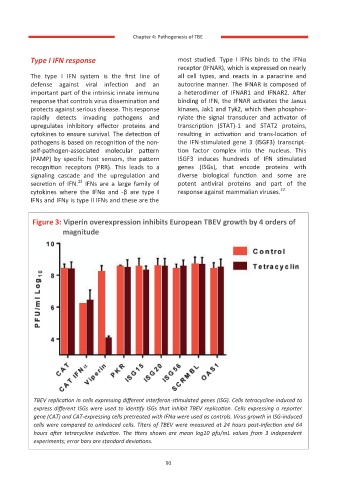
           Figure 3: Viperin overexpression inhibits European TBEV growth by 4 orders of
                    magnitude


           TBEV replication in cells expressing different interferon-stimulated genes (ISG). Cells tetracycline-induced to
           express different ISGs were used to identify ISGs that inhibit TBEV replication. Cells expressing a reporter
           gene (CAT) and CAT-expressing cells pretreated with IFNα were used as controls. Virus growth in ISG-induced
           cells were compared to uninduced cells. Titers of TBEV were measured at 24 hours post-infection and 64
           hours  after  tetracycline  induction.  The  titers  shown  are  mean  log10  pfu/mL  values  from  3  independent
           experiments; error bars are standard deviations.


                                                     90

