


Page 154 - TBE_Book_V2_2019
P. 154
Chapter 8: TBE in animals


          system  to  eliminate  the  virus  within  a  few      Figure 6
          days  is  probably  the  reason  for  a  fatal
          outcome, as in most of these cases no specific
          intrathecal  antibody  production  and  no
          increased cell count in the CSF were detected
                      17
          prior to death.
          CSF analysis in affected dogs with clinical signs
          mostly reveals elevated leukocyte count, with
          predominantly   mononuclear   cells   and
          elevated  total  protein.  CSF  changes  are
          concomitant  to  virus  elimination  and  rising
          antibody  titers.  Specific  antibodies  are
          detectable  in  the  serum  of  affected  dogs
          within  a  few  days. 7,17,18,20   Cross-reactivity  to
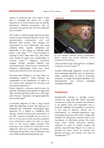
          Louping  ill  virus,  West  Nile  virus,  and  Usutu   An  old  Labrador  Retriever  during  rehabilitation.
          virus  should  be  taken  into  consideration  in   Water  training  over  months  improved  muscle
                        10,23                        strength and coordination.
          endemic  areas.    Magnetic  resonance
          imaging  findings  included  bilateral  and   observed when dogs reach partial or complete
          symmetrical gray matter lesions involving the   remission of clinical signs. 17,26
          thalamus,  hippocampus,  brain  stem,  basal
          nuclei, and ventral horn on the spinal cord.    Possible  differential  diagnoses  include  other
                                                      viral  meningoencephalitis  such  as  distemper,
          All lesions had minimal or no mass effect, or   rabies,  pseudo-rabies,  as  well  as  protozoal,
                           24
          perilesional  edema.   These  findings  are   bacterial,  or  fungal  meningoencephalitis,  and
          comparable  to  the  distribution  of  lesions  in   paraneoplastic   and   immune   mediated
          the  canine  brain  detected  by  necropsy  and   meningoencephalitis.
                             25
          immunohistochemistry.
          Proton  magnetic  resonance  spectroscopy,  to
          evaluate metabolic abnormalities in dogs with   Treatment
          TBE, revealed significant differences with dogs
          with  immune  mediated  meningoencephalitis
                        26
          and healthy dogs.                           Symptomatic  therapy  is  strongly  recom-
                                                      mended  for  dogs  with  TBE.  Water  and  food
                                                      maintenance orally, by constant rate infusion,
          A  tentative  diagnosis  of  TBE  in  dogs  should   or  by  gastric  tubes  and  supportive  care  is
          fulfill  the  following  criteria:  tick  exposure  or
          observed  tick  infestation,  neurological  signs   essential. Sedation and relaxation is necessary
                                                      in  the  case  of  seizures.  Steroid  use  is
          indicative  for  a  diffuse  or  multifocal  CNS   controversial,  as  immune-suppression  may
          disease,  (mostly  mononuclear)  pleocytosis  in   prolong the presence of the virus. In dogs with
          the CSF, a positive antibody titer in serum or   marked  CSF  pleocytosis,  steroids  seem  to  be
          CSF, or in the case of fatal outcome a positive
          virus  confirmation  within  the  brain  or  spinal   mandatory  to  effectively  protect  the  brain
                                                      tissue  from  further  fulminant  immune
          cord.  In  the  future,  highly  sensitive  PCR   response.  In  cases  of  muscle  atrophy  and
          techniques may include virus detection in the   paresis,  physiotherapy  (Figure  6)  as  early  as
          diagnostic work-up in early stages of disease.
                                                      possible  has  been  shown  to  improve  the
          Increasing serum titers may be detected, but
                                                      general  outcome  and  shorten  the  time  of
          more often rapidly decreasing titers are               19,20
                                                      rehabilitation.

                                                  149
                                                  149

