
Page 26 - TBE_Book_V2_2019
P. 26
Chapter 2a: Virology
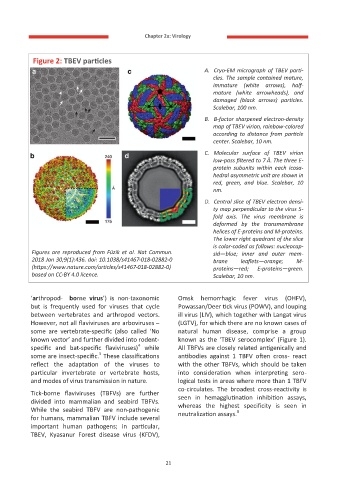
Figure 2: TBEV particles
A. Cryo-EM micrograph of TBEV parti-
cles. The sample contained mature,
immature (white arrows), half-
mature (white arrowheads), and
damaged (black arrows) particles.
Scalebar, 100 nm.
B. B-factor sharpened electron-density
map of TBEV virion, rainbow-colored
according to distance from particle
center. Scalebar, 10 nm.
C. Molecular surface of TBEV virion
low-pass filtered to 7 Å. The three E-
protein subunits within each icosa-
hedral asymmetric unit are shown in
red, green, and blue. Scalebar, 10
nm.
D. Central slice of TBEV electron densi-
ty map perpendicular to the virus 5-
fold axis. The virus membrane is
deformed by the transmembrane
helices of E-proteins and M-proteins.
The lower right quadrant of the slice
is color-coded as follows: nucleocap-
Figures are reproduced from Füzik et al. Nat Commun. sid—blue; inner and outer mem-
2018 Jan 30;9(1):436. doi: 10.1038/s41467-018-02882-0 brane leaflets—orange; M-
(https://www.nature.com/articles/s41467-018-02882-0) proteins—red; E-proteins—green.
based on CC-BY 4.0 licence. Scalebar, 10 nm.
‘arthropod- borne virus’) is non-taxonomic Omsk hemorrhagic fever virus (OHFV),
but is frequently used for viruses that cycle Powassan/Deer tick virus (POWV), and louping
between vertebrates and arthropod vectors. ill virus (LIV), which together with Langat virus
However, not all flaviviruses are arboviruses – (LGTV), for which there are no known cases of
some are vertebrate-specific (also called ‘No natural human disease, comprise a group
known vector’ and further divided into rodent- known as the ‘TBEV serocomplex’ (Figure 1).
4
specific and bat-specific flaviviruses) while All TBFVs are closely related antigenically and
5
some are insect-specific. These classifications antibodies against 1 TBFV often cross- react
reflect the adaptation of the viruses to with the other TBFVs, which should be taken
particular invertebrate or vertebrate hosts, into consideration when interpreting sero-
and modes of virus transmission in nature. logical tests in areas where more than 1 TBFV
co-circulates. The broadest cross-reactivity is
Tick-borne flaviviruses (TBFVs) are further seen in hemagglutination inhibition assays,
divided into mammalian and seabird TBFVs. whereas the highest specificity is seen in
While the seabird TBFV are non-pathogenic 6
neutralization assays.
for humans, mammalian TBFV include several
important human pathogens; in particular,
TBEV, Kyasanur Forest disease virus (KFDV),
21