
Page 30 - TBE_Book_V2_2019
P. 30
Chapter 2a: Virology
protein molecules are located at their N- and proteins rather than by the nucleocapsid
C-termini and are separated by hydrophobic protein.
regions. The nucleocapsid is less ordered and In addition to mature virions, smaller (approxi-
as for other flaviviruses, no discernible mately 14 nm in diameter) non-infectious
symmetry was detected in cryoelectron particles are released from the infected cells.
26
microscopic reconstructions. Instead, the C
These particles lack nucleocapsid and consist
protein is arranged in a cage-like structure of E and M proteins only; they are called
surrounding the viral genome. The icosahedral sedimenting (70S) hemagglutinin (SHA).
symmetry is, therefore, directed by surface
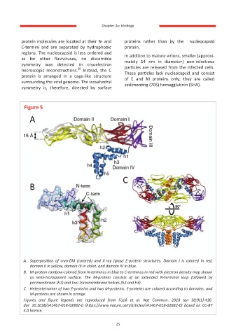
Figure 5
A. Superposition of cryo-EM (colored) and X-ray (gray) E-protein structures. Domain I is colored in red,
domain II in yellow, domain III in violet, and domain IV in blue.
B. M-protein rainbow-colored from N-terminus in blue to C-terminus in red with electron density map shown
as semi-transparent surface. The M-protein consists of an extended N-terminal loop followed by
perimembrane (h1) and two transmembrane helices (h2 and h3).
C. Heterotetramer of two E-proteins and two M-proteins. E-proteins are colored according to domains, and
M-proteins are shown in orange.
Figures and figure legends are reproduced from Füzik et al. Nat Commun. 2018 Jan 30;9(1):436.
doi: 10.1038/s41467-018-02882-0 (https://www.nature.com/articles/s41467-018-02882-0) based on CC-BY
4.0 licence.
25