
Page 27 - TBE_Book_V2_2019
P. 27
Chapter 2a: Virology
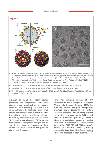
Figure 3
A. Schematic model of a flavivirus particle. Left panel: immature virion, right panel: mature virion. The surface
of immature particles consists of 60 spikes composed of trimers of prM-E heterodimers. Mature particles are
formed after prM cleavage and contain 90 E homodimers. (From Vratskikh O, Stiasny K, Zlatkovic J, et al.
Dissection of antibody specificities induced by yellow fever vaccination. PLoS Pathog 2013;9:e1003458.
figshare: https://dx.doi.org/10.1371/journal.ppat.1003458.g001 (CC BY)).
B. Pseudoatomic cryo-EM reconstruction model of the immature flavivirus particle (PDB: 2OF6).
C. Pseudoatomic cryo-EM reconstruction model of the mature flavivirus particle (PDB: 3J0B).
D. Cryo-EM micrograph of immature TBEV particles (kindly provided by Tibor Füzik and Pavel Plevka, with per-
mission). Scalebar, 100 nm.
Although all TBFVs are closely related Three main antigenic subtypes of TBEV
genetically and antigenically, they cause correspond to the 3 recognized genotypes:
diverse clinical manifestations in humans: Western, also known as European (TBEV-EU;
OHFV and KFDV (including a subtype of this previously Central European encephalitis;
virus, Alkhurma hemorrhagic fever virus) prototype strain Neudoerfl), Far Eastern
induce hemorrhagic fever syndromes, while (TBEV-FE; previously Russian spring-summer
the others cause neurological disease. encephalitis; prototype strain Sofjin), and
Importantly, the hemorrhagic fever-associated Siberian (TBEV-Sib; previously Western
TBFVs and encephalitogenic TBFVs do not Siberian encephalitis; prototype strains
form separate phylogenetic lineages and no Zausaev and Vasilchenko). 10,11 Two additional
specific determinants in the genomes of these lineages; i.e., “178-79” and “886-84 group”,
viruses have been associated with particular named as Baikalian TBEV (TBEV-Bkl)
7,8
disease manifestations. respectively, have been identified in Eastern
Siberia and proposed as TBEV subtypes. 116, 117
22