


Page 119 - TBE_Book_V2_2019
P. 119
Chapter 5: TBE in adults


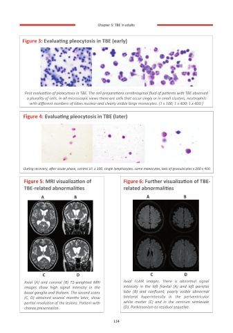
           Figure 3: Evaluating pleocytosis in TBE (early)


            First evaluation of pleocytosis in TBE. The cell preparations cerebrospinal fluid of patients with TBE observed
             a plurality of cells. In all microscopic views there are cells that occur singly or in small clusters, neutrophils
              with different numbers of lobes nuclear and clearly visible large monocytes. (1 x 100; 1 x 400; 1 x 400.)

           Figure 4: Evaluating pleocytosis in TBE (later)


           During recovery, after acute phase, control LP. x 100, single lymphocytes, some monocytes, lack of granulocytes x 200 x 400.


           Figure 5: MRI visualization of             Figure 6: Further visualization of TBE-
           TBE-related abnormalities                  related abnormalities

                   A                 B                             A             B


                   C              D                                 C                    D
           Axial  (A)  and  coronal  (B)  T2-weighted  MRI   Axial  FLAIR  images.  There  is  abnormal  signal
           images  show  high  signal  intensity  in  the   intensity  in  the  left  frontal  (A)  and  left  parietal
           basal ganglia and thalami. The second scans   lobe  (B)  and  confluent,  poorly  visible  abnormal
           (C, D) obtained several months later, show   bilateral  hyperintensity  in  the  periventricular
           partial resolution of the lesions. Patient with   white  matter  (C)  and  in  the  centrum  semiovale
           chorea presentation.                       (D). Parkinsonism as residual sequelae.

                                                  114
                                                  114

